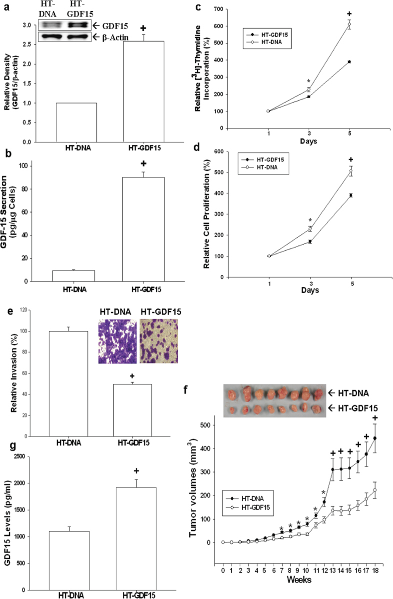
Figure 3. Effect of GDF15 overexpressed on cell proliferation, invasion, and tumorigenesis in HT1376 cells.
Expressions of GDF15 in HT1376 cells stably transfected with pcDNA3 (HT-DNA) or pcDNA-GDF15 (HT-GDF15) expression vector were determined by immunoblotting assays (a) and ELISA (b). Data are expressed as mean-fold (±S.E.; n = 3) in relation to the HT-DNA cell group and the mean (±S.E.; n = 6) of the GDF15 levels. Proliferations of HT-DNA (white circle) and HT-GDF15 (black circle) cells were determined according to the incorporation of 3H-thymidine (c) and MTS assays (d). Each point on the curve represents the mean-percentage (±S.E.; n = 6) of that on day 1. (e) The invasive ability of cells was determined by in vitro matrigel invasion assays. Data are presented as mean-percentage (±S.E.; n = 3) in relation to that of the HT-DNA cell group. (f) Nude mice were inoculated subcutaneously with HT-DNA (black circle) or HT-GDF15 (white circle) cells. At the indicated days, tumor size was measured using vernier calipers, and results are presented as tumor size in mm3 (±S.E.). (g) Blood samples were collected from experimental animals by cardiocentesis immediately after sacrificed, and GDF15 levels were determined by ELISA. Data is presented as mean (±S.E.; n = 6) of the GDF15 levels. (*P < 0.05, +P < 0.01).

