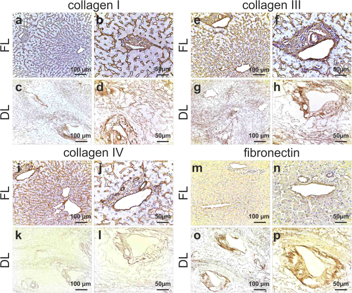
Figure 3. Expression and distribution of ECM proteins.
Collagen I, III and IV staining in FL is seen as fine strands in the parenchymal space as well as around the blood vessels (a,b; e,f; I,j). Collagen I and III distribution was preserved following decellularization as demonstrated by a staining in both sinusoids and portal tracts in DL (c,d; g,h). Collagen IV (k,l) and fibronectin (o,p) staining showed a conserved meshwork in sinusoids and biliary ducts after decellularization. Scale bar for 20X magnification (left panel): 100 μm and 40X (right panel): 50 μm.

