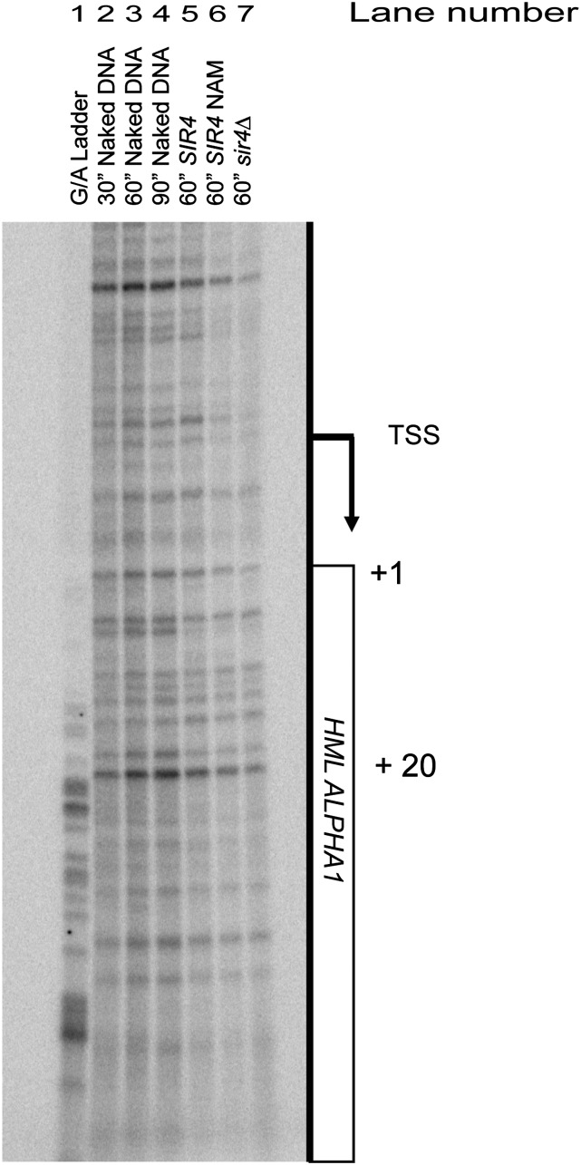
Figure 1.
The KMnO4 reactivity of HMLα1 in vivo. The pattern of KMnO4 reactivity is shown for the promoter and 5′ region of HMLα1 coding region. Genomic sequences of A+G are shown as a G/A ladder in lane 1. Naked genomic DNA reacted with 20 mM KMnO4 for various times is shown in lanes 2–4. The pattern of reactivity of this region in cells reacted with KMnO4 is shown in lanes 5–7. The reactivity pattern for cells with HMLα repressed lane 5, cells in which HML was derepressed with 5 mM nicotinamide (lane 6) or genetically by sir4∆ (lane 7) all lack enhanced cleavage sites characteristic of paused/stalled RNA polymerase. The arrow denotes the transcription start site (TSS) (Zhang and Dietrich 2005). The numbers on the right side of the panel identify bases in HMLα1 beginning at the initiation ATG codon as +1.

