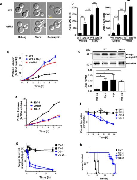
Figure 2. A Vad1 RCK/p54 Homolog Plays a Role in Repression of Autophagy and Virulence in C. neoformans.

(a, b) Indicated strains were observed in mid-log phase, starvation in phosphate-buffered saline (PBS), pH 7.4 (starv) or in the presence of rapamycin by differential interference contrast microscopy (DIC) for the presence of autophagic bodies (AB, white arrows) and quantified in 200 cells per condition by DIC in n=3 independent experiments. Chi square analysis: *** p < 0.001.
(c) Autophagic flux determined by 35S pulse-labeling of cells followed by precipitation in 10% tricholoroacetic acid (TCA) at indicated times followed by measurement of TCA-soluble peptides by liquid scintillography.. Autophagic flux was determined for the indicated strains in mid-log phase (wild type [WT], Δvad1) or in mid-log cells in the presence of rapamycin (+Rap) for indicated times. Time points n=3 +/− SEM.
(d) Lysate of mid-log phase cells either treated (+) or untreated (−) with rapamycin or after starvation in PBS, pH 7.4 (starv) were subjected to urea electrophoresis and detected on western blot using an anti-Atg8 rabbit antibody. Lower arrow indicates lipidated Atg8 bands. Lower panel represents results of n=3 independent experiments +/− SD. * p < 0.05; ** p < 0.01
(e) Autophagic flux in starved cells was determined as in (a). C. neoformans expressing empty vector strain #1 (EV-1) and VAD1 overexpressing vector, strain #1 (OE-1). Time points n=3 +/− SEM.
(f) Fungal cell survival under starvation conditions. Strains were starved by incubation in PBS, pH 7.4, for the indicated times, followed by live cell determination using an assay of colony-forming units (CFU). C. neoformans expressing empty vector strain #1 and #2 (EV-1 and 2) and VAD1 overexpressing vector (under the ACT1 promoter), strain #1 and 2 (OE-1 and 2). Time points n=3 +/− SE.
(g) Fungal survival in macrophages. Indicated strains were opsonized with 40% mouse serum, phagocytosed using a J774.16 cell line induced by IFNG/IFN-γ, and fungal viability measured by CFU. Time points n=3 +/− SEM.
(h) Mouse mortality after cryptococcal infection. Indicated strains (1 × 10⁶ CFU) were inoculated intravenously into 10 female Swiss-Albino mice for each strain and followed until moribund. n=10 mice per group.
