
Figure 6. DDX6 is a Suppressor of Autophagy in HeLa cells.
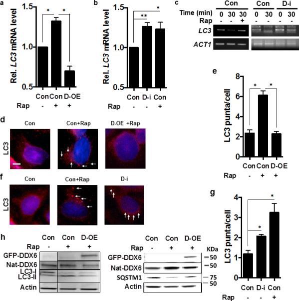
(a-b) LC3 transcript levels in HeLa cells in nutrient-rich media, transformed with empty vector (con) or empty vector + rapamycin (Con+Rap) or with a plasmid expressing human DDX6 in the presence of rapamcyin (D-OE + Rap) or treated with non-targeted siRNA (Con), or DDX6-directed siRNA (D-i), or non-targeted siRNA in the presence of rapamycin (Con+Rap). N=3 independent experiments +/− SD, Student t-tes; * p <0.05, ** p <0.01.
(c) The presence of capped LC3 transcripts was assayed in HeLa cells subjected to DDX6-directed siRNA (DDX6-i) or non-targeted siRNA (Con) under the indicated conditions at the indicated times after transcriptional suppression by the specific PCR method detailed in Methods.
(d-e) HeLa cells subjected to conditions in Fig. 3a and examined by fluorescence microscopy for LC3 puncta formation using an LC3-II antibody (red fluorescence, white arrows; 4',6-diamidino-2-phenylindole used for nuclear localization (blue fluorescence) and quantified in n=3 independent experiments of 100 cells each +/− SD. Scale bar = 2 μm. Student's t-test, * p < 0.05.
(f-g) HeLa cells subjected to conditions in Fig. 3b, and examined by fluorescence microscopy for LC3 puncta formation using anLC3-II antibody (red fluorescence, white arrows; 4',6-diamidino-2-phenylindole used for nuclear localization (blue fluorescence) and quantified in n=3 independent experiments of 100 cells each +/− SD; Student's t-test; * p < 0.05.
(h) HeLa cells in conditions equivalent to Fig. 3a were assayed for the indicated proteins including cells containing plasmids expressing GFP-DDX6 and native DDX6 (Nat-DDX6) by western blot according to Methods.