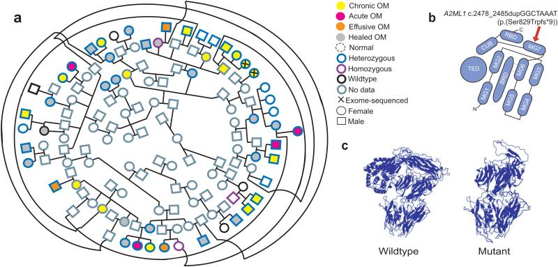
Figure 1. Segregation within the indigenous pedigree, cartoon of A2ML1 domains and molecular modeling for the A2ML1 variant.
[A] Pedigree connecting 37 variant carriers who have the full spectrum of otitis media (age 3 months to 58 years, median 13 years). An individual with healed otitis media and intellectual disability (ID) is wildtype for the duplication, but her 13-year old son who has chronic otitis media but no ID and her unaffected 4-month old daughter are heterozygous. Nine variant carriers had no evidence of otitis media (median 18 years), while four individuals are wildtype and unaffected. [B] Predicted A2ML1 domains based on alpha 2-macroglobulin structure.9 MG, macroglobulin-like domains 1-7; BRD, bait-region domain; CUB, consists of two four-stranded antiparallel β-sheets; TED, thiol-ester binding domain; RBD, receptor-binding domain. The A2ML1 frameshift variant is expected to occur within the MG7 domain (red arrow). [C] Modeling predicts loss of the receptor-binding and thiol-ester domains due to the A2ML1 duplication.

