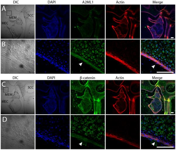
Figure 2. A2ML1 is localized to middle ear epithelium.
[A, C] Sagittal cryosections from P6 wildtype mice showing the middle ear cavity (MEC), the middle ear mucosa (MEM) and semicircular canal of the inner ear (SCC). [B, D] Higher magnification depicting boxed sections in A and C. [A-D] Confocal images of the cryosections immunostained with antibodies for DAPI (blue), A2ML1 (green in A & B), β-catenin (green in C & D) and rhodamine phalloidin (Actin, red). White arrowheads point to MEM with A2ML1 [B] and β-catenin expression [D]. DIC, differential interference contrast. Scale bar: 100 μm.

