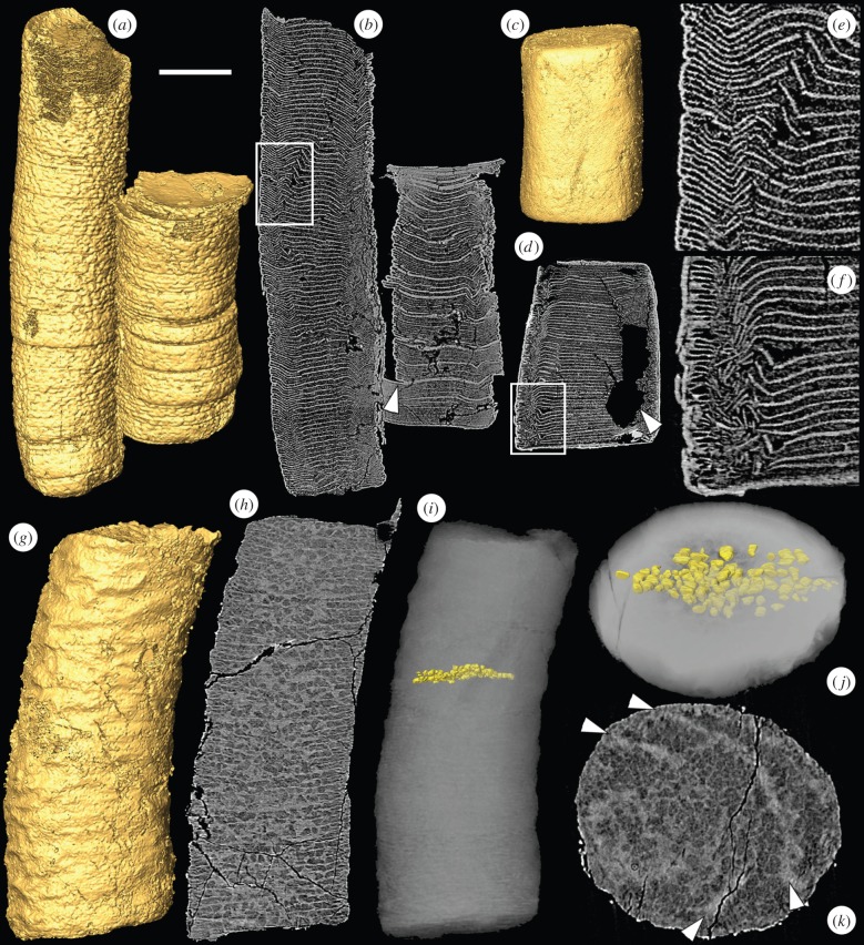
Figure 1.
SRXTM images of the tubular fossils Sinocyclocyclicus and Quadratitubus. (a) SRXTM surface model of Sinocyclocyclicus, X 5322; (b) longitudinal SRXTM slice through the specimen in (a) showing brittle deformation of the cross walls, the arrowhead indicates displacement along the plane of the cross wall, the region in the box is enlarged in (e); (c) surface model of Quadratitubus, X 5323; (d) longitudinal SRXTM section through the specimen in (c) showing brittle deformation, the arrowhead indicates a void created by pyrite dissolution, the region in the box is enlarged in (f); (e) higher magnification image of boxed region in (b); (f) higher magnification image of boxed region in (d); (g) SRXTM surface model of a specimen of Sinocyclocyclicus with ovoid to sub-cuboid structures preserved in the spaces between the cross walls, X 5324; (h) longitudinal SRXTM slice of the specimen in (g); (i) reconstruction based on PXCT data showing the arrangement of structures between two successive cross walls; (j) reconstruction in (i) shown in oblique view; (k) transverse SRXTM slice of the specimen in (g), the arrowheads indicate the positions of two successive cross walls. Scale bars: (a,b) 120 µm; (c,d) 143 µm; (e) 38 µm; (f) 40 µm; (g–i) 58 µm; (j,k) 32 µm. (Online version in colour.)

