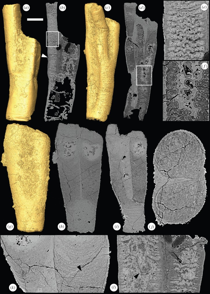
Figure 2.
SRXTM images of the tubular fossil Ramitubus. (a) SRXTM surface model of Ramitubus, X 5325, showing ductile deformation of the tube; (b) longitudinal SRXTM section through the specimen in (a) showing ductile deformation of the crosswalls, the arrowhead indicates a region where there are several successive compartments of lower than average height, the region in the box is enlarged in (e); (c) SRXTM surface model of Ramitubus, X 5326; (d) longitudinal SRXTM section through the specimen in (c) showing organic degradation of the cross walls, the region in the box is enlarged in (f); (e) higher magnification image of boxed region in (b); (f) higher magnification image of boxed region in (d) showing cross walls truncated by an irregular region of void-filling cement; (g) SRXTM surface model of Ramitubus, X 5327; (h) longitudinal SRXTM section through the specimen in (g), note the high-attenuation (bright), nearly spherical structures towards the end of each branch; (i) longitudinal section through the specimen in (g) at a different level, note the change in the pattern of the cross walls after branching; (j) transverse SRXTM section through the specimen in (g); (k) higher magnification SRXTM section of the specimen in (g) showing finer layers between the cross walls, the arrowhead shows a region where these layers are made up of sub-angular structures; (l) longitudinal SRXTM section through the Ramitubus specimen in (a), the arrowhead indicates down-folding of a cross wall that results in a prominent pocket-like space towards the centre of one of the specimen. Scale bars: (a,b) 183 µm; (c,d) 126 µm; (e) 48 µm; (f) 40 µm; (g–i) 89 µm; (j) 36 µm; (k) 36 µm; (l) 64 µm. (Online version in colour.)

