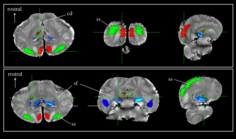
Figure 2.
Thalamic parcellation of Delphinus delphis based on cortical regions. Three regions of interest were defined: (i) visual cortex (red); (ii) auditory cortex based on traditional boundaries in the suprasylvian gyrus (green) and (iii) auditory region temporal cortex based on IC tractography (blue). Seeds within the entire thalamus were traced to these regions and thresholded above 20 000 streamlines. Each row shows a set of slices that correspond to a single cursor location. The upper row is located posteriorly through the cortex while the lower row is located through thalamus. The thalamic parcellations to the three regions demonstrate that the temporal region is primarily connected to the ventrocaudal region, presumably near the medial geniculate nucleus. The visual and auditory regions overlap substantially in the thalamus and are located more dorsally and rostrally.

