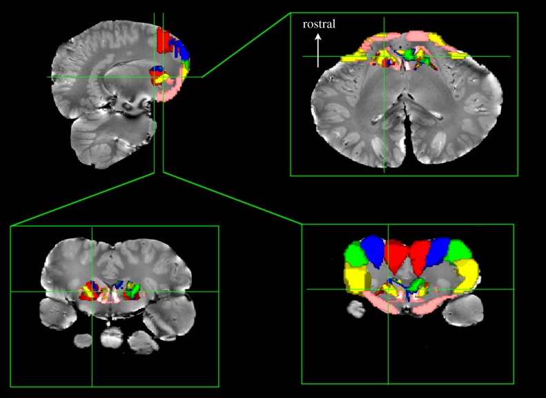
Figure 4.
Basal ganglia parcellation based on cortical regions in Delphinus delphis. Five regions were defined in the orbital (‘frontal’) lobes from the longitudinal fissure (red) progressing laterally and ventrally (blue, green and yellow) to the ventral orbital lobe (pink). Seeds within the entire basal ganglia were traced to these regions and thresholded above 20 000 streamlines. Bottom row shows two coronal slices through different parts of the basal ganglia. The dorsal portions of the basal ganglia are connected to the dorsal portions of the cortex (red, blue and green), while the ventral basal ganglia are connected to the ventrolateral (yellow) and ventral orbital lobe (pink).

