
Figure 1.
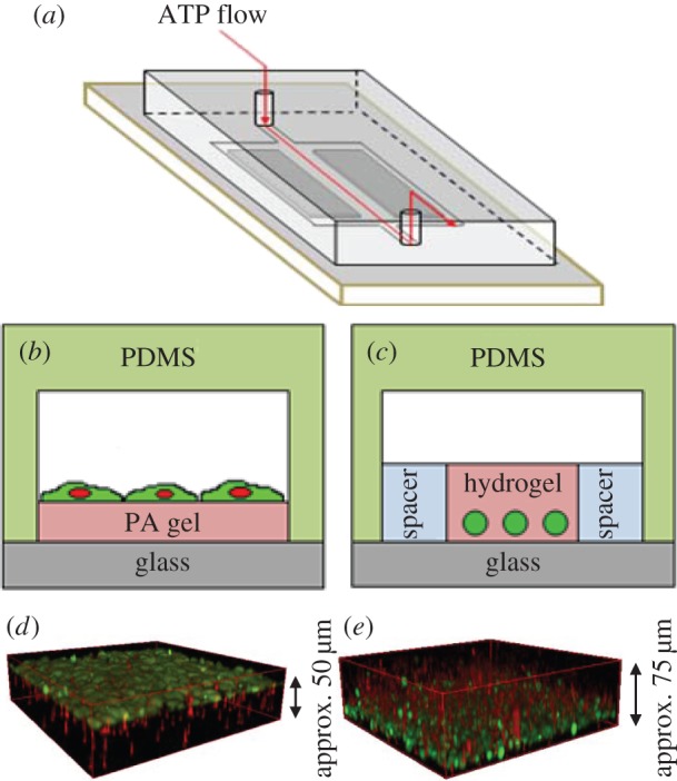
Experimental set-up used in chemosensing experiments with fibroblast cells. (a) Schematic of the flow device. (b,c) Cross-sectional view of two-dimensional and three-dimensional chemosensing experiments, respectively. In the two-dimensional set-up, cells (green) are cultured on top of polyacryamide (PA) gels with various values of Young's modulus and are directly in contact with ATP solution. In the three-dimensional set-up, cells are encapsulated in hydrogel with various values of Young's modulus. ATP flows on top and diffuses through the gel, exciting the cells located on the bottom. (d,e) Confocal z-stack three-dimensional reconstruction of fibroblast cells on top of PA gel (d) and inside the hydrogel (e). Red fluorescence represents the polystyrene beads embedded in the polyacrylamide gel or hydrogel, and green fluorescence represents the fibroblast cells. (Online version in colour.)