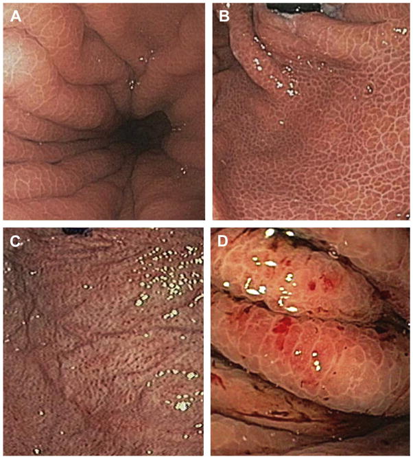
Fig. 1.
(A, B) Representative images of mild PHG. A shows a forward-viewing image of the proximal stomach. B shows a retroflex view of the cardia with the classic form of PHG, the typical “mosaic-like pattern” without significant stigmata of bleeding or erythema or edema. (C, D) Representative images of severe PHG. Red lesions of variable diameter are evident. There is often irregular mucosa. Cherry spots may be confluent or not. Slow oozing may also be seen as in D, an up-close view in the proximal stomach.

