
Figure 5.
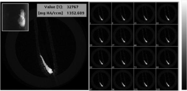
X-ray microCT imaging of PEG-Cu2S NCs at tube voltage of 45 KeV. Cross sectional X-ray contrast imaging of phantom with PEG-Cu2S NCs (left). The inset in the left shows the guide view of the same. Z-stacked contrast image of the PEG-Cu2S NCs is shown in right with the intensity scale.