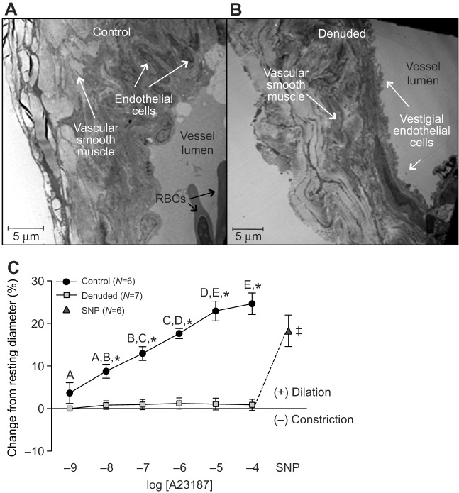
Fig. 2.
Trout coronary microvessel structure before and after endothelial denudation, and vasomotor responses to increasing concentrations of the calcium ionophore A23187. Denuded vessels were exposed to CHAPS (0.4%) by intraluminal injection (0.3 ml) for approximately 2 min. Intact endothelial cells are visible on the luminal surface of the control vessel (A), while in the treated vessel the endothelial cell layer is visibly reduced (B). See Materials and methods for details on TEM methodology. (C) Vasomotor responses of trout coronary microvessels to increasing concentrations of A23187. SNP (sodium nitroprusside, 10−4 mol l−1) was administered at the end of each trial to test for vascular smooth muscle viability in the denuded vessels. ‡ indicates a significant difference between denuded microvessels after exposure to 10−4 mol l−1 of A23187 versus SNP injection. Dissimilar letters indicate significant differences between concentrations of A23187, *P<0.05 between control (intact) and denuded vessels at a particular concentration of A23187. Note that the series of sham injections (saline containing ethanol) did not result in any noticeable (i.e. ≤0.5%) changes in vessel ID. Thus, we considered any change in vessel ID greater than 2% (mean change plus 2 s.d. caused by sham injection) to be biologically significant. All concentrations are in mol l−1.

