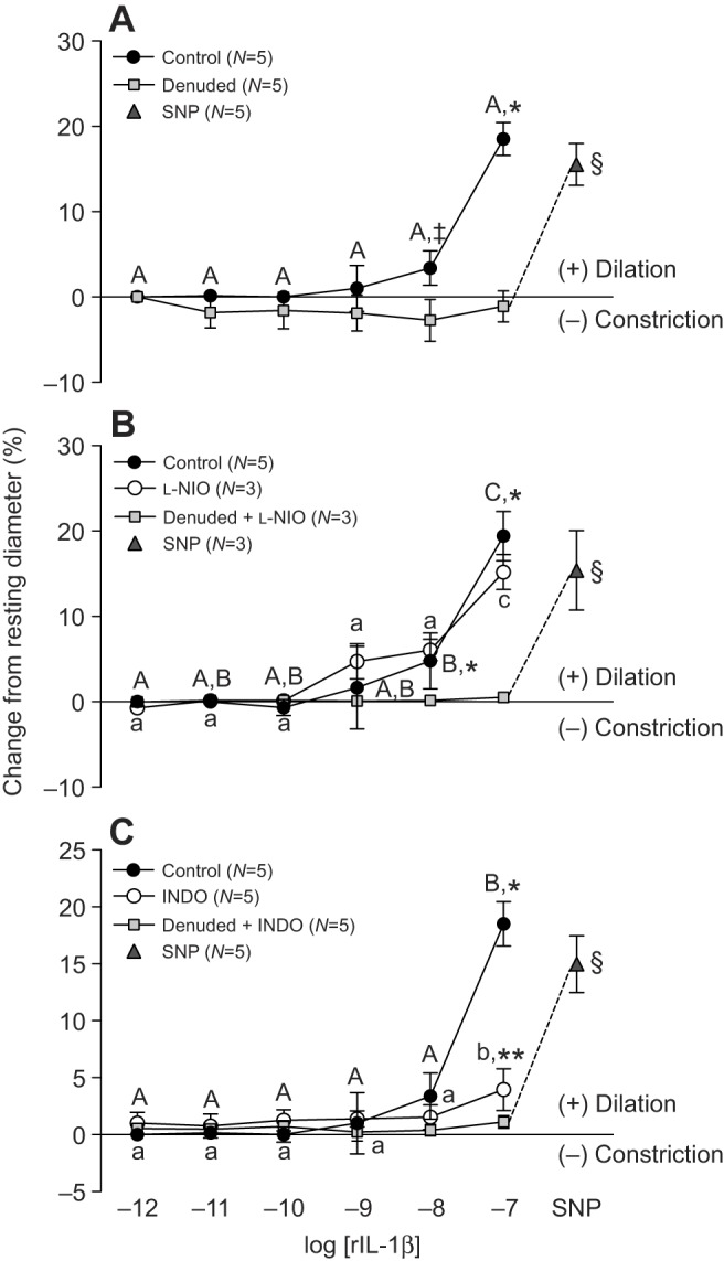
Fig. 3.

Effect of vessel denudation, l-NIO and indomethacin on trout coronary microvessel dilation in response to recombinant interleukin-1β. (A) Vessel denudation was achieved by exposing the intraluminal surface of the vessel to the non-ionic detergent CHAPS (0.4%). Vessels were incubated for 30 min with either the NOS inhibitor L-NIO (10−4 mol l−1) (B), or the COX inhibitor INDO (10−4 mol l−1) (C), before rIL-1β injections were initiated. Dissimilar capital letters indicate significant (P<0.05) differences in the response to the various concentrations of rIL-1β in control (intact) vessels, while dissimilar lower case letters indicate differences in the response to rIL-1β in vessels incubated with either L-NIO (B) or INDO (C). Significant differences between control (intact) and denuded vessels are indicated by *, while ** indicates a significant difference between vessels in the presence (open circles) versus absence (filled circles) of blocker (i.e. L-NIO or INDO) at a particular concentration of rIL-1β (P<0.05, except where indicated ‡, where 0.1>P≥0.05). Sodium nitroprusside (SNP, 10−4 mol l−1) was administered at the end of each trial to test for vascular smooth muscle viability in denuded vessels, and § indicates a significant difference between denuded microvessels after exposure to 10−7 mol l−1 rIL-1β (with or without blocker) versus after SNP injection. Note that a series of sham (saline containing DTT) injections did not result in any noticeable (i.e. ≤1%) change in vessel ID. Thus, we considered any change in vessel ID greater than 3% (mean change plus 2 s.d. caused by the sham saline injections) to be biologically significant. All concentrations are in mol l−1.
