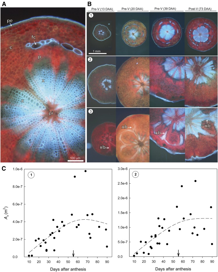
Figure 2.
Free-hand cross sections of pedicels imaged unstained under violet fluorescent light over fruit development. Lignified tissue appears in bright blue color. A, Enlarged image of the pedicel stem portion indicating the presence of various tissue types. c, Cortex; fc, fiber cap cells; p, phloem; pe, periderm, pi, pith; r, less lignified ray cells; x, lignified xylem tissue. B, Spatial and temporal changes in pedicel dimensions and xylem cross-sectional area of stem portion (see row 1, position 1 in Fig. 10) and receptacle portion (see rows 2 and 3, positions 2 and 3 in Fig. 10, respectively). In the receptacle portion close to the fruit, radial branches of xylem tissue (xb) into the cortex are visible (row 3), which increase in intensity over fruit development. V, Veraison. C, Developmental changes in Ax in stem portion (image 1, left) and receptacle portion (image 2, right). Dashed lines indicate 75% smoothed trend lines. The arrows indicate veraison.

