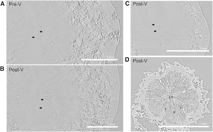
Figure 5.
Representative transverse microCT images through the pedicel showing water-filled tissue in light gray and air-filled tissue in dark gray. Black arrows indicate some air-filled tissue in the pith. A and B, Receptacle portion (i.e. position 2 in Fig. 10) of pedicels scanned preveraison at 34 DAA (A) and postveraison at 76 DAA (B). C, Stem portion scanned at 76 DAA (i.e. position 1 in Fig. 10). These images were acquired from pedicels that were scanned immediately after they were cut under water from fruit and rachis. D, Stem portion of a dehydrated pedicel scanned at 85 DAA. c, Cortex; pi, pith; r, ray cells; x, xylem tissue.

