Figure 6.
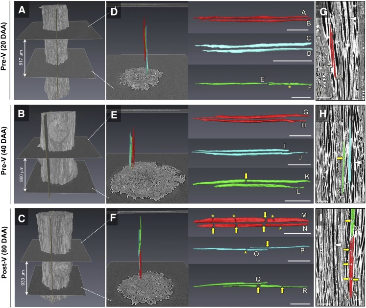
Three-dimensional (3-D) reconstructions of xylem vessel elements in the pedicel stem portion at 20, 40, and 80 DAA. Pedicels were scanned after dehydration. A to C, Volume renderings of the pedicel surface. D to F, Reconstructed vessel elements (n = 6 each) within that pedicel portion. Images to the right show enlarged versions of these reconstructed vessel elements. For better visualization, pairs of vessel elements that were located in close proximity to each other are given the same color (i.e. red, turquoise, or green). G to I, 2-D images of reconstructed vessel elements as they are embedded in the pedicel apoplast (light gray; air-filled vessel lumen is shown in dark gray, as indicated by white arrowheads). The positions of the longitudinal 2-D image slices are indicated in A to C. c, Cortex; pi, pith. Asterisks indicate constricted vessel lumen. Yellow arrows indicate positions where the continuity of the vessel lumen is interrupted. Bars = 100 μm.

