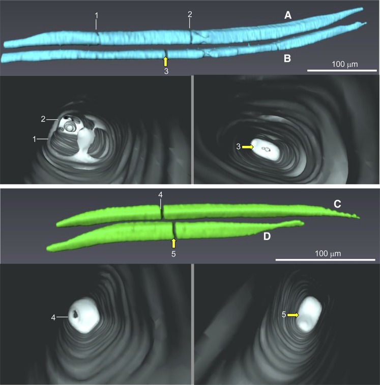
Figure 7.
3-D reconstructions of xylem vessel elements in the pedicel stem portion at 85 DAA (i.e. postveraison). Images were obtained from a dehydrated pedicel scanned at high resolution. At positions where vessel elements are constricted (A at positions 1 and 2 and C at position 4) or interrupted (B at position 3 and D at position 5), the deposition of some material is visualized inside the vessel lumen. Yellow arrows indicate the positions of blockages in the lumen of vessel elements.

