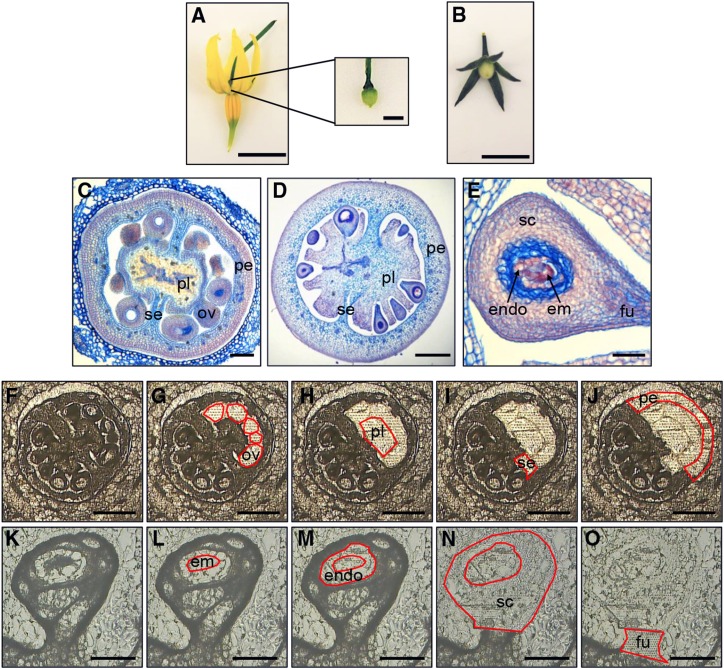
Figure 1.
LCM of S. pimpinellifolium ovaries and fruit. A and B, Photographs of flower and ovary (A) at 0 DPA and fruit (B) at 4 DPA. Bars = 5 mm and 500 μm (inset). C to E, Transverse paraffin sections of 0-DPA ovaries (C), 4-DPA fruit (D), and 4-DPA seeds (E). Labels indicate ovules (ov), placenta (pl), septum (se), pericarp (pe), endosperm (endo), embryo (em), seed coat (sc), and funiculus (fu). Bars = 50 μm (C and E) and 300 μm (D). F to J, LCM of ovaries at 0 DPA. Cryosections show a whole ovary before (F) and after collection of ovules (G), placenta (H), septum (I), and pericarp (J). Bars = 300 μm. K to O, Cryosections of seed from a 4-DPA fruit before (K) and after removal of embryo (L), endosperm (M), seed coat (N), and funiculus (O). Bars = 100 μm.

