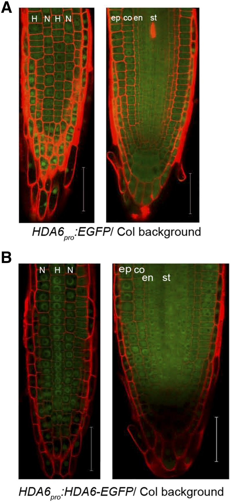
Figure 2.

HDA6 localizes ubiquitously in Arabidopsis root tip. A, Confocal laser scanning microscope (CLSM) images of 7-d-old root tip of HDA6pro:EGFP reporting the location of HDA6 expression: epidermal view (left) and median view (right). Bars = 50 μm. B, CLSM images of 7-d-old root tip of HDA6pro:HDA6-EGFP showing HDA6 protein localization: epidermal view (left) and median view (right). Bars = 50 μm. In all images, N indicates nonhair cell position and H indicates hair cell position. co, Cortex; en, endodermis; ep, epidermis; st, stele. Red areas are propidium iodide (PI) signals and green areas are GFP signals.
