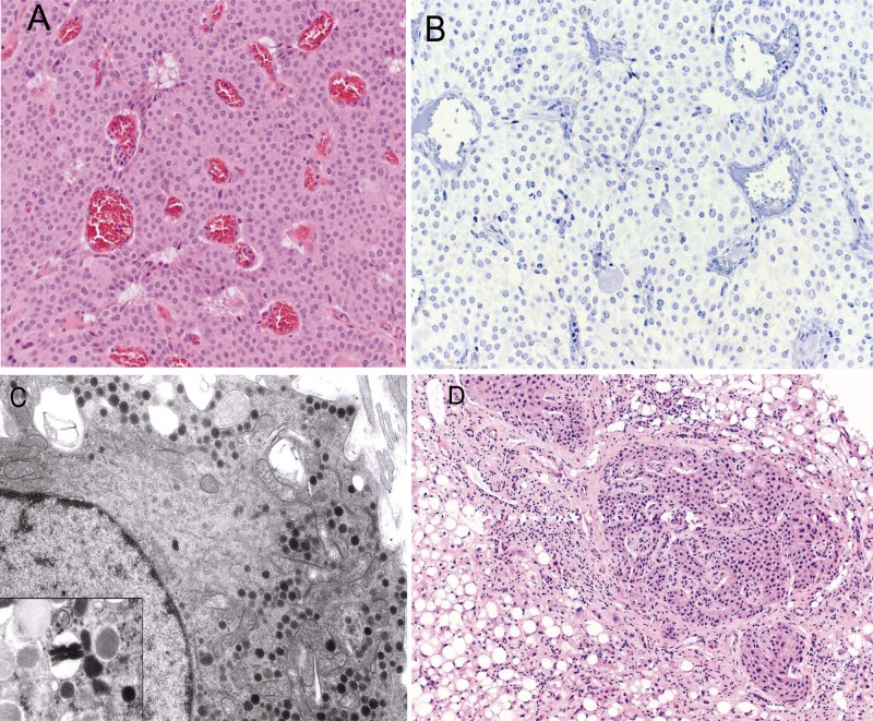
Figure 2.
(A) Pancreatic tumor histology. Representative hematoxylin and eosin–stained section of resected pancreatic mass showed a well-differentiated endocrine tumor (original magnification, 20×). (B) Immunohistochemical staining for insulin was negative (original magnification, 20×). (C) Electron micrograph of pancreatic tumor. The cytoplasm contained round neuroendocrine granules with dense cores; none of the granules had the typical crystalloid structure of insulin granules (original magnification, 6000×); inset: example of crystalloid insulin granules from a normal islet (original magnification, 20 000×). (Images courtesy Elizabeth Sengupta, MD, and Jerome Taxy, MD, The University of Chicago Medical Center.) (D) Biopsy of liver lesion showed a well-differentiated endocrine neoplasm (hematoxylin and eosin stain; original magnification, 10×)

