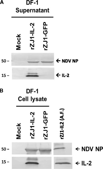
Fig. 1.

Production of chicken IL-2 was assessed by western blot in vitro (DF-1 cell line) and in vivo (infected eggs) at 48 hpi. A strong band of approximately 15 kDa is present in blots from the supernatant (panel a), cell lysate (panel b), and allantoic fluid (panel b) of rZJ1-IL2-infected DF-1 cells and eggs. No band of the same molecular weight was present in blots from the rZJ1-GFP-infected groups. Blots from the supernatant (panel a), cell lysates (panel b), and allantoic (panel b) fluid of both rZJ1-IL2 and rZJ1-GFP groups showed bands for NDV NP at approximately 50 kDa. No bands were observed in mock-inoculated controls, either at 15 or 50 kDa
