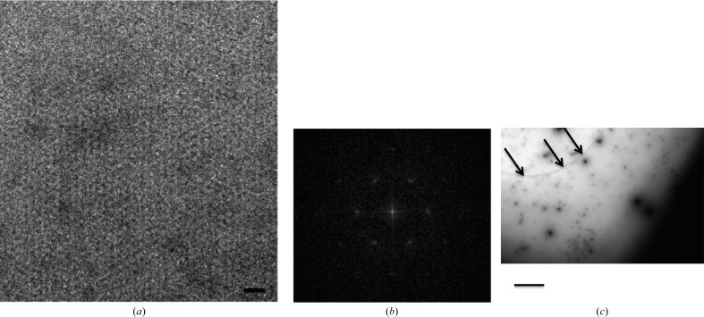
Figure 1.
Electron-microscopic characterization of two-dimensional crystals of ABCG2. (a) Negative-stain electron-microscopic image of two-dimensional crystals of ABCG2 with 2%(w/v) uranyl acetate. The scale bar represents 35 nm. (b) A respective power spectrum. (c) An overview of the crystal at lower magnification, where the arrows show where the crystal originates. The scale bar represents 500 nm.

