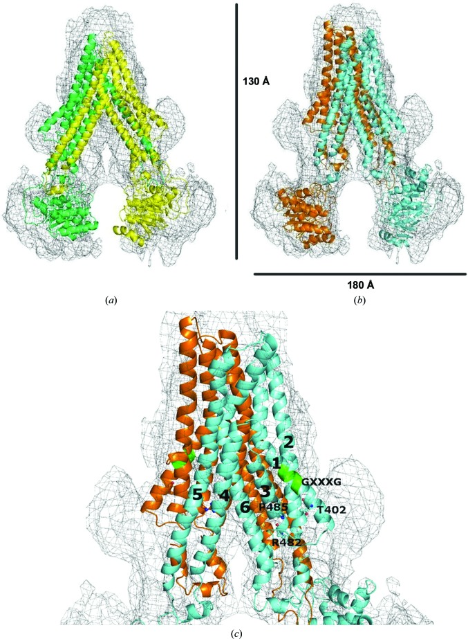
Figure 6.
Alignment of homology models to the ABCG2 map. (a) Fitting of the original closed apo inward-facing model to the map. (b) Fitting of the refined inward-facing model to the map. (c) A closer look at the TMDs. TM α-helices in the refined model are labelled 1, 2, 3, 4, 5 and 6 for TM1, TM2, TM3, TM4, TM5 and TM6 of one ABCG2 monomer (blue), respectively. Residues Thr402 (T402), Arg482 (R482) and Pro485 (P485) as well as the GXXXG motif are depicted near their locations in one ABCG2 monomer (blue). The three residues are indicated in ball-and stick representation and the GXXXG motif in TM1 is shown in green.

