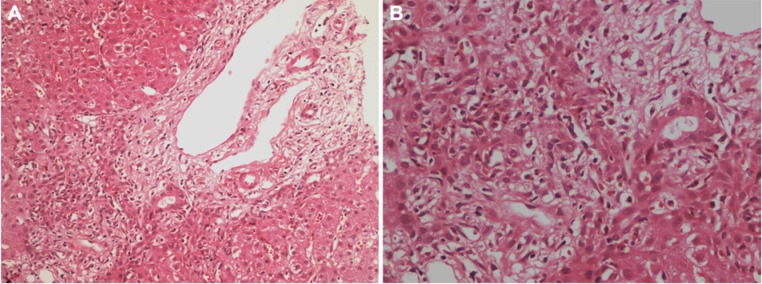
Figure 1.

(A) Fibrous portal expansion, bile duct proliferation, and a mixed inflammatory infiltrate predominantly consisting of polymorphonuclear leukocytes, partially disrupting the limiting plate are seen (hematoxylin–eosin; magnification 200×). (B) Mixed inflammatory reaction in portal tract and parenchyma, predominantly consisting of polymorphonuclear leukocytes; number of eosinophils, few lymphocytes, and plasmocytes are also seen in this field (hematoxylin–eosin; magnification 400×).
