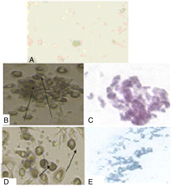
Figure 1.

Morphological and histological staining of differentiated MSCs/BM. A. Undifferentiated MSCs. B. Differentiated MSC osteoblasts after addition of growth factors. C. MSCs differentiated into osteoblasts stained with Alizarin red. D. Arrows for differentiated MSC chondrocytes after addition of growth factors. E. MSCs differentiated into chondrocytes stained with Alcian blue. MSCs/BM, bone marrow-derived mesenchymal stem cells.
