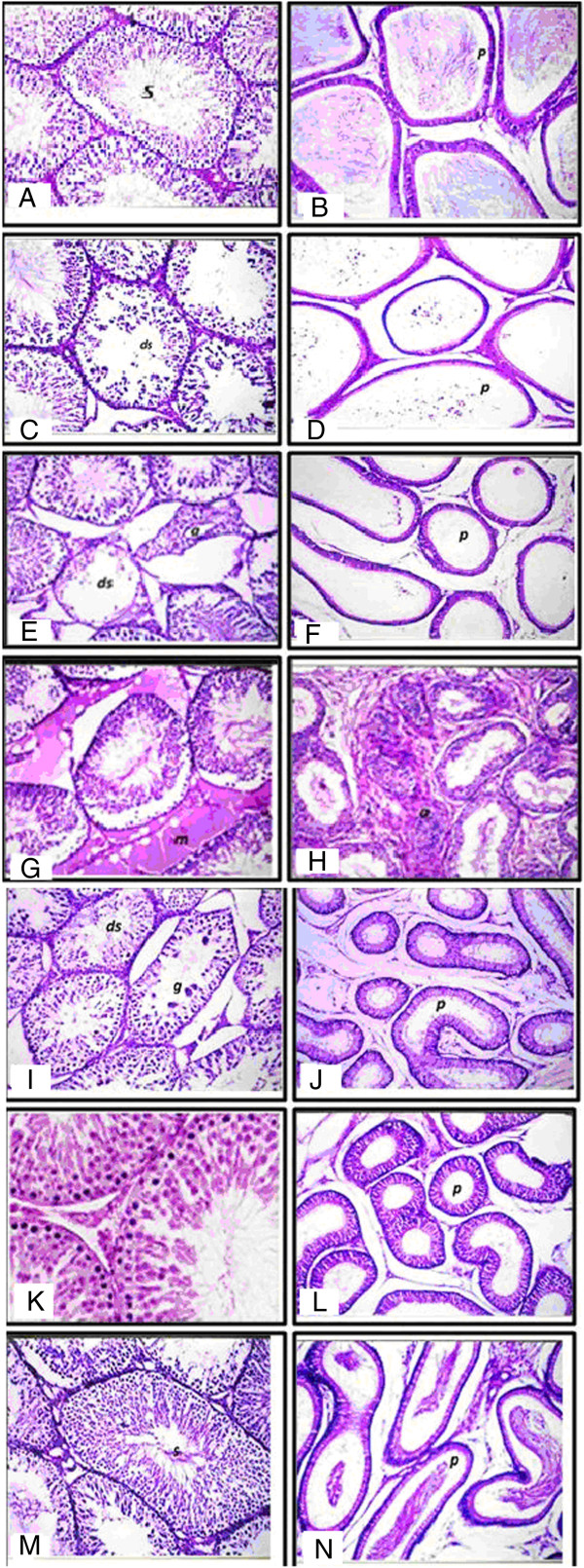
Figure 8.

Histopathology showing the alterations induced by LN and treatment effects of MSCs/BM in testis tissues of rat. Photomicrographs of H & E-stained sections of (A): testes of control rats showing normal histological structure of the mature active seminiferous tubules with complete spermatogenic sense (s) × 40. B: epididymis of rats in the control group showing normal histological structure of the tubules and impacted by mature sperm (p) × 40. C: photomicrographs of H & E stained sections of LN groups after 21 days showing degeneration in some seminiferous tubules (ds) × 40. D: epididymus of rats in the LN group post 21 days showing epididymal tubules lumen (p). E and F: section of testes of rat in the LN group post 30 days showing degeneration (ds) and atrophy (a) of some individual seminiferous tubules (8E), and epididymal tubules free from sperm (p) (8 F). G and H: section of the testes rat post 60 days of LN showing homogenous eosinophilic structure material replacing the interstitial of Leydig cells (m) (8G) and anaplastic activity in the lining epithelium of the epididymal tubules (a) with empty lumen (8H). I and J: photomicrographs of testes post 21 days of MSCs/BM showing degeneration (ds) with giant spermatogonial formation (g) of some seminiferous tubules (8I) and epididymal tubular lumen (p) with swelling in the lining epithelium × 40 (8 J). K and L: section of testes post 30 days of MSCs/BM treatment showing mild degeneration in some seminiferous tubules (ds) (8 K) with spermatogonial formation (8 L). M and N: testes of rat post 60 days of MSCs/BM treatment showing normal intact histological structure with complete active seminiferous tubules sense (s) (8 M) and intact normal histological structure of the epididymal tubules impacted by spermatozoa in the lumen (p) × 40 (8 N). LN, lead nitrate; MSCs/BM, bone marrow-derived mesenchymal stem cells.
