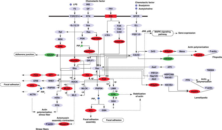
Figure 6. Proteins involved in the regulation of actin cytoskeleton.

KEGG pathway analysis of proteins differentially phosphorylated by IL-33 using DAVID functional analysis tool indicated the enrichment of the Regulation of actin cytoskeleton pathway. Proteins identified to be regulated by IL-33 are represented in red (hyperphosphorylated) or green (hypophosphorylated).
