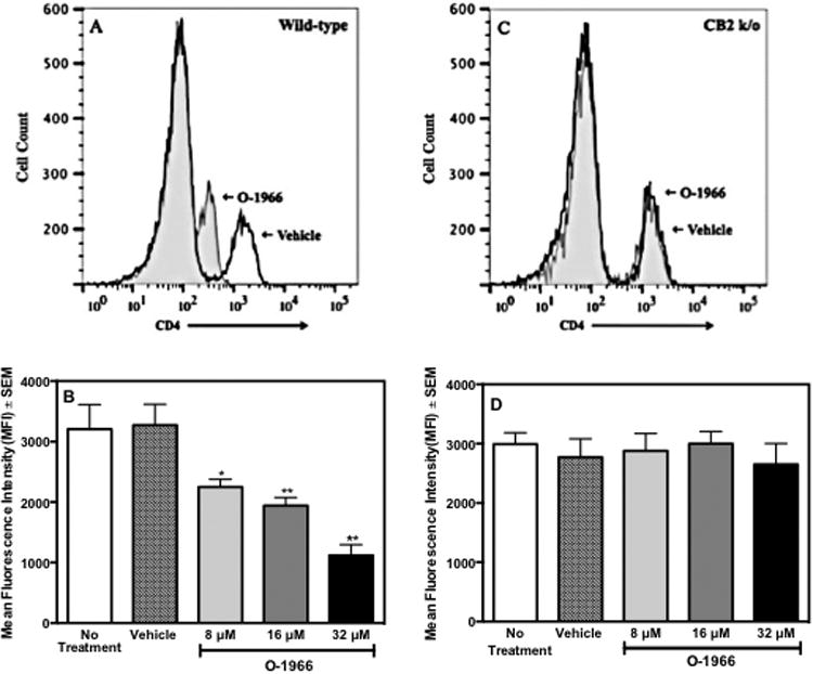
Figure 4.

O-1966 treatment decreases CD4 expression in vitro. Wild-type (panels A and B) or CB2R k/o (panels C and D) C57BL/6 responder splenocytes were pretreated for 3 h with O-1966 or ethanol vehicle. MLR cultures were harvested at 48 hr and analyzed for CD4 expression on CD3+CD4+ populations by flow cytometry. An equal number of CD3+CD4+ cells were analyzed for each treatment group. Representative histograms of CD3+ cells from cultures treated with 32 μM O-1966 (gray filled) compared to vehicle treated cells (white filled) with responder cells from wild-type (Panel A) and CB2R k/o (Panel C). Mean Fluorescence Intensity (MFI) of CD4 in CD3+CD4+populations from MLR cultures that received no treatment (
 ), ethanol vehicle (
 ), 8 μM O-1966 (
 ), 16 μM O-1966 (
 ), or 32 μM O-1966 (
 ). Data are mean ± S.E.M. of 3 separate experiments. *p < 0.01, **p < 0.001 (ANOVA, O-1966 versus vehicle). Values for vehicle are not significantly different from no treatment.
