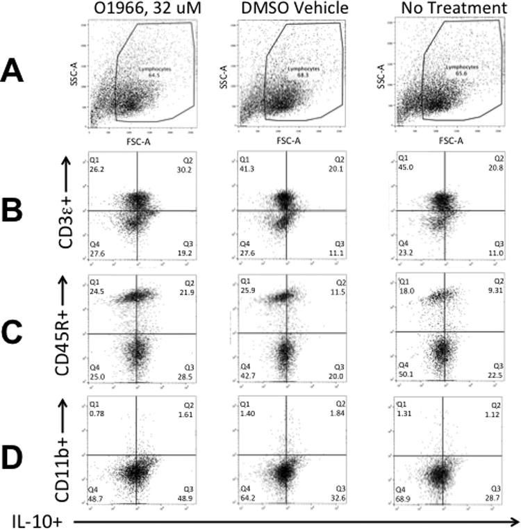
Figure 8.

Detection of cell subsets expressing intracellular IL-10 in MLR cultures treated with O1966. Cells were first stained with eFluor 780 Fixable Viability Dye (eBioscience), then with antibodies to outer surface markers, and then stained intracellularly with APC-labeled anti-mouse IL-10. Row A: Plot of forward scatter (FSC-A) vs. side scatter (SSC-A), showing gating for lymphocytes of O-1966 treated cells, DMSO treated cells, and no treatment cells. Row B: Plot of eFluor 450 CD3e+ (T-cells) vs. IL-10+ cells. Row C: Plot of PE-Cy7 CD45R+(B220+) (B-cells) vs. IL-10+ cells. Row D: Plot of BV605 CD11b+ (macrophages) vs. IL-10+ cells. The quandrants indicate the percentage of live cells expressing both IL-10 and the indicated surface marker shown on the Y-axis. Quadrants were set using unstained cells.
