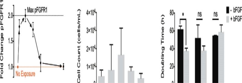
Fig. 1.
Primary human myoblast exposure to bFGF results in cellular responses that occur over a range of time scales from 100 – 103 min. (A) Phosphorylation of FGFR1 (pFGFR1) in primary myoblasts peaks 5 min after bFGF stimulation. (B) Stimulation with 5 ng/mL bFGF promoted the greatest amount of migration in myoblasts as measured in Transwell assays over 6 h. Error bars represent the standard deviation between three independent studies. (C) The doubling time for myoblasts with or without bFGF stimulation was determined on laminin, fibronectin, and collagen. Error bars represent standard deviation; * p < 0.05 and ns = not significant (p > 0.05).

