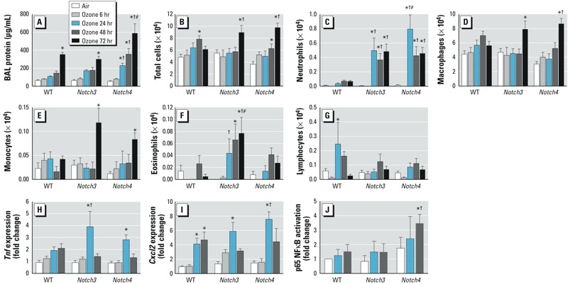
Figure 1.

Notch3–/– and Notch4–/– mice are more susceptible to ozone-induced lung inflammation compared with WT mice. (A) Bronchoalveolar lavage fluid (BALF) protein concentration increased significantly after 72 hr ozone in all mice; protein concentration increased earliest (24 hr) and most significantly in Notch4–/– mice. (B) BALF total cells increased in WT mice after 48 hr and in Notch3–/– and Notch4–/– mice after 72 hr. (C) Neutrophils significantly increased in Notch3–/– and Notch4–/– mice after 24–72 hr compared with air-exposed and WT mice. BAL macrophages (D) and monocytes (E) significantly increased in Notch3–/– and Notch4–/– mice after 72 hr. (F) BAL eosinophils significantly increased only in Notch3–/– mice. (G) BAL lymphocytes significantly increased only in WT mice after 24 hr. (H) Expression of Tnf in whole-lung homogenates significantly increased in Notch3–/– and Notch4–/– mice after 24 hr. (I) Expression of Cxcl2 in whole-lung homogenates increased in all groups after 24 hr; n = 4–10 mice per group (exposed in 2–3 groups). In A–I, *p < 0.05 compared with respective air controls, †p < 0.05 compared with WT at same time point, and #p < 0.05 between Notch3–/– and Notch4–/– mice at the same time point, by 2-way ANOVA with Bonferroni post hoc tests. (J) p65 NF-κB activation was significantly increased after 48 hr ozone in Notch4–/– mice and when compared with WT and Notch3–/– mice. *p < 0.05 compared with respective air control, and †p < 0.05 compared with WT and Notch3–/– ozone, by 2-way ANOVA with Holm-Sidak pairwise comparisons.
