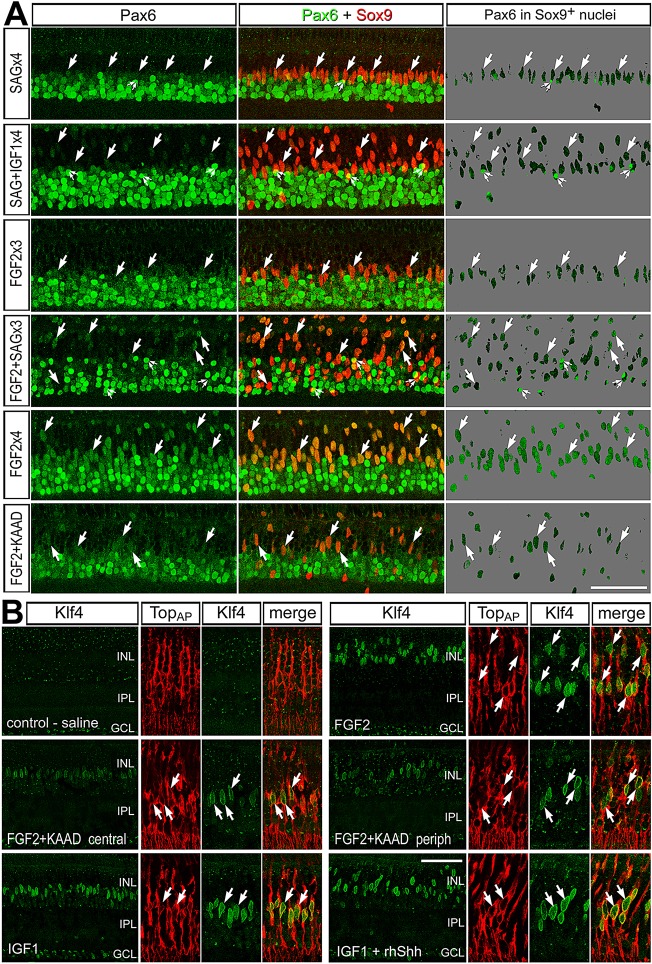
Fig. 7.
Müller glia and MGPCs express Pax6 and Klf4 in response to treatment with different growth factors and/or Hh agonists/antagonists. (A,B) Retinas were treated with consecutive daily doses of saline ×4, SAG ×4, SAG and IGF1 ×4, FGF2 ×3, FGF2 and SAG ×3, FGF2 ×4 or FGF2 and KAAD-cyclopamine ×4. Retinas were immunolabeled for Pax6 (green; A) and Sox9 (red; A), or Klf4 (green; B) and TopAP (red; B). Arrows indicate the nuclei of Müller glia and/or MGPCs. Images were obtained by confocal microscopy, projecting four optical sections; therefore, there was some overlap of adjacent amacrine and glial nuclei through the z axis, indicated by small double arrows. GCL, ganglion cell layer; INL, inner nuclear layer; IPL, inner plexiform layer; ONL, outer nuclear layer. Scale bars: 50 µm.

