Fig. 8.
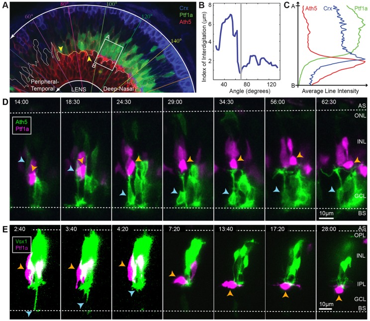
Early stages of IPL formation. (A) Snapshot of a 55 hpf retina showing different stages of retinal development along the developmental wave. The RIN labelled with the orange asterisk probably represents a misplaced cell. (B) Quantification of the degree of interdigitation along the apical surface of RGCs, drawn as a white line in A. The vertical grey line indicates the angular position of the minimum value, and the corresponding 20° segment is indicated by the yellow line and yellow arrowheads in A. (C) The average line intensity values of the boxed region in A, showing the BC plexus (Crx) located between the RGC (Ath5) dendritic plexus and RINs (Ptf1a) in this region. (D) Selected frames of a movie of an RGC (blue arrowheads) migrating basally past RINs (orange arrowheads) and stratifying at a location basal to the RINs. (E) Selected frames of a movie of a BC axon (blue arrowheads) retracting to the basal side of RINs in the INL before the subsequent migration of a dAC (orange arrowheads) into the GCL. Time is shown as h:min from the start of the imaging sessions, at ∼44 hpf (D) and ∼48 hpf (E).

