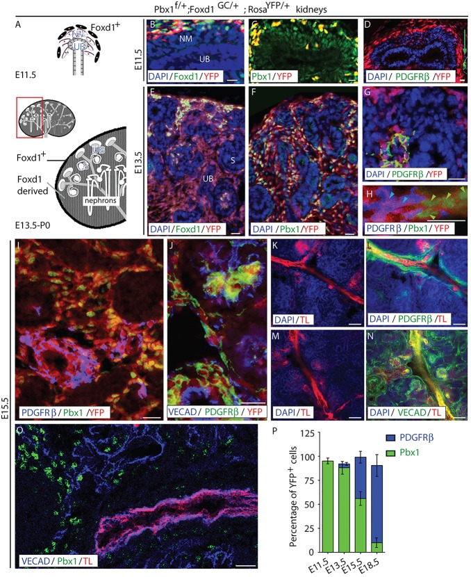
Fig. 1.
Pbx1 is expressed by VMC progenitors prior to their differentiation. (A) Diagram of the tissues forming the developing kidney at the initiation of kidney development (E11.5, top) and at later stages of development (E13.5-P0, bottom). The E11.5 kidney rudiment includes renal epithelial progenitors, including the ureteric bud (UB), nephrogenic mesenchyme (NM) and peripheral Foxd1+ mesenchyme, which gives rise to VMCs. Later in development, a population of self-renewing Foxd1+ VMC-progenitors persists at the periphery of the kidney, whereas Foxd1-derived progeny are located within the interstitial compartment between developing S-shaped body and more mature nephrons. (B-J) Frozen sections of kidneys from Pbx1f/+; Foxd1GC/+; RosaYFP/+ embryos in which YFP is expressed in all cells derived from Foxd1+-VMC progenitors. E11.5 (B-D), E13.5 (E-H) and E15.5 (I,J) renal tissues were assayed by IF for the detection of Foxd1 (B,E), Pbx1 (C,F,H,I), PDGFRβ (D,G,H,I,J) and VE-cadherin (VECAD; J). The self-renewing VMC progenitor population was identified by co-expression of Foxd1 and YFP fluorescence, VMC progenitors by YFP fluorescence, and definitive Foxd1-derived VMCs by PDGFRβ and YFP expression. VECAD staining was used to visualize vascular endothelia. At E11.5 (B), the vast majority of Foxd1+ progenitors located at the periphery of the rudiment express the YFP lineage tag (D) and Pbx1 (C). Differentiated VMCs, as determined by PDGFRβ+ expression, are not present in the kidney at this stage, although they can be seen in extra-renal tissues (D, green arrow). At E13.5, cells co-expressing Foxd1+ and Pbx1 remain localized to the periphery of the rudiment (E), whereas their Pbx1+ progeny, which have downregulated Foxd1, are present in the interstitial compartment (F). At this stage, small populations of lineage-tagged cells expressing PDGFRβ are observed (G,H). These differentiated VMCs lack Pbx1 (H; green arrowheads indicate Pbx1+YFP+ nuclei; light blue arrowheads indicate PDGFRβ+YFP+ VMCs). At E15.5, large populations of differentiated PDGFRβ+Pbx1− lineage-tagged VMCs are observed (I). These cells are localized around blood vessels (J), further confirming their VMC phenotype. Scale bars: 10 μm. (K-O) E15.5 kidneys processed by intravital dye labeling using Tomato Lectin (TL) to label blood-conducting vessels (K-O) and subsequent IF detection of PDGFRβ (L), VECAD (N,O) and Pbx1 (O). PDGFRβ+ VMCs are selectively associated with mature, blood-conducting vessels, whereas the majority of VECAD+ cords and vessels are associated with VMC progenitors that have yet to differentiate. Scale bars: 50 μm. (P) Quantification of the percentage of YFP+ Foxd1 progeny expressing Pbx1 or PDGFRβ at different stages of development (n≥3 genotype/stage). Error bars represent s.d. See also supplementary material Fig. S1.

