
Fig. 7.
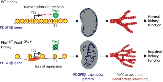
Model for Pbx1-dependent vascular patterning in the developing kidney. In wild-type (WT) kidney, during the initial stages of renal development, Pbx1 binds the E1 cis-regulatory element of Pdgfrb, repressing transcription and spatially restricting its expression to cortical domains of the kidney. As a result, the temporal and spatial association of VMCs with the developing vasculature is tightly regulated leading to the normal hierarchical pattern of the renal arterial network required for renal function. By contrast, in Pbx1f/f;Foxd1GC/+ kidneys, Pdgfrb repression is released in the VMC progenitors. This de-repression results in precocious and ectopic Pdgfrb expression in VMCs, the premature association of PDGFRβ+ VMCs with the nascent vascular bed, abnormal arterial branching, impaired renal function and neonatal demise.