Figure 1.
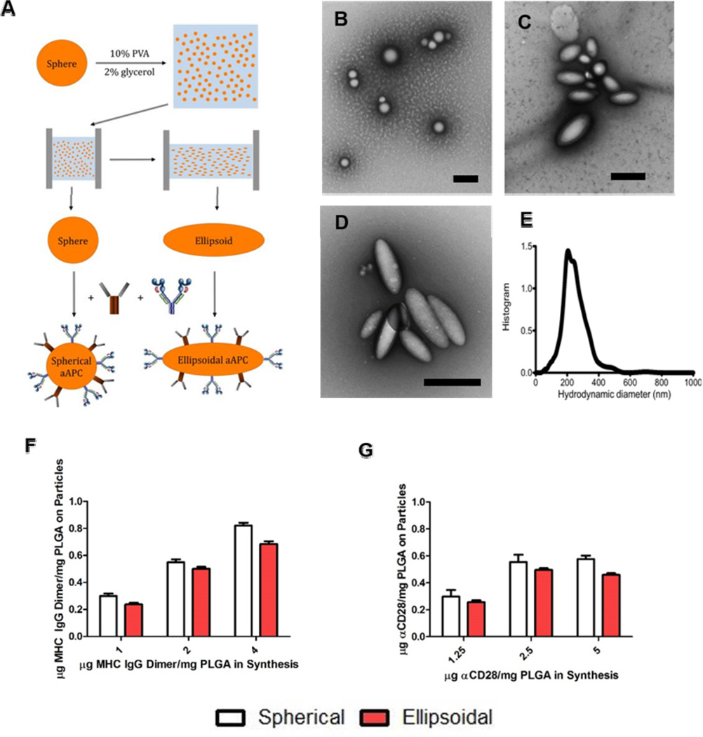
Non-spherical and spherical nanodimensional artificial antigen presenting cell (naAPC) characterization. (a) PLGA nanoparticles were synthesized by single emulsion and elongated utilizing the film stretching method. Conjugation of MHC Db Ig Dimer and anti CD-28 mediated by EDC/NHS chemistry resulted in naAPCs. (b,c,d) TEM images of (b) non-stretched spherical particles (c) 2-fold stretched particles, and (d) 3-fold stretched particles. Scale bars are 500 nm. (e) Particles were sized by Nanoparticle Tracking Analysis and determined to be 224 nm in size. The particle protein conjugation efficiency on spherical and 2-fold stretched ellipsoids for (f) MHC Db Ig dimer and (g) anti CD-28 was analyzed by conjugation of fluorescent protein. Conjugation results demonstrate similar amounts of protein bound to each particle shape. Error bars represent standard errors of >3 trials.

