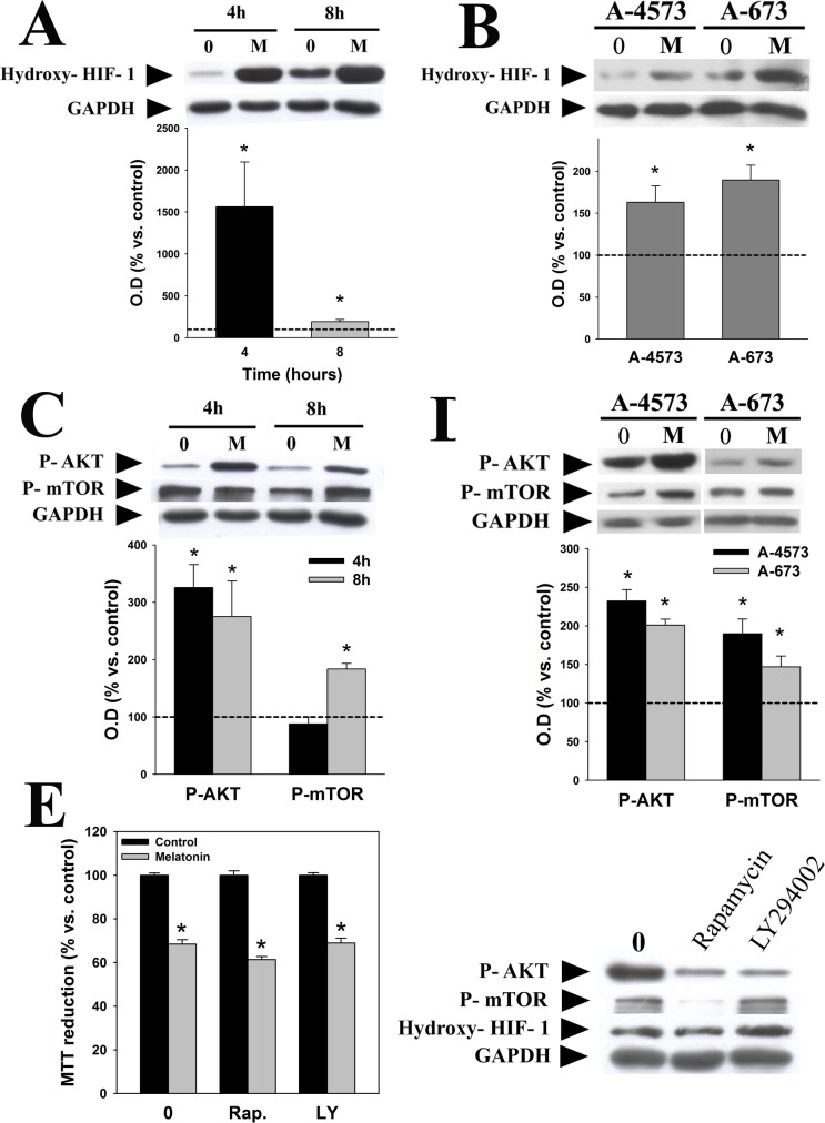
Fig 5. Melatonin inhibits HIF-1α in a PI3K/AKT/mTOR independent pathway.
Western blot analyses were carried out to identify the effect of melatonin (1 mM, 4 and 8 hours) on the activation of HIF-1α in TC-71 cells (A) and A-4573 and A-673 (1mM, 4 hours) cells (B). AKT and mTOR activation in TC-71 (C) and A-4573 and A-673 cells (D) was evaluated using specific phosphoantibodies. GAPDH was used as a loading control in all cases. A representative blot is showed. Optical density of bands was measured and values of the hydroxy-HIF-1α (inactivated form), p-AKT or p-mTOR bands were normalized versus GAPDH. Results are represented as percentage of the values found in vehicle-treated cells (dotted line). (E) Left panel, cell viability was evaluated by MTT reduction assay after treatment of TC-71 cells with 1 mM melatonin alone or in combination with 10 nM rapamycin or 10 μM LY294002 for 48 hours. Data are expressed as the percentage of vehicle-treated cells. Right panel, Representative western blot showing the relative protein level of p-AKT, p-mTOR and hydroxy-HIF-1α after 10 nM rapamycin or 10 μM LY294002 treatment during 24 hours in TC-71 cell line. p*≤0.05 vs. vehicle-treated cells.

