Figure1.

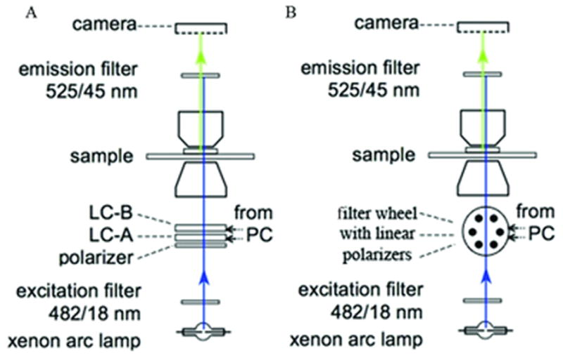
Schematic of polarized fluorescence microscope light path. Light path set up for polarized fluorescence imaging using an LC (A) or linear polarizers (B). Adapted from DeMay et al., 2011b.

Schematic of polarized fluorescence microscope light path. Light path set up for polarized fluorescence imaging using an LC (A) or linear polarizers (B). Adapted from DeMay et al., 2011b.