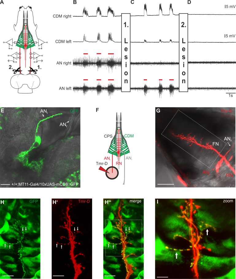
Fig 4. Motor neurons for pharyngeal pumping innervate the cibarial dilator musculature (CDM) ipsi- and contra-laterally.
A, Simultaneous antennal nerve (AN) and cibarial dilator muscle (CDM) recordings of left and right side of the larval body of an OregonR larva. B, Under unimpaired conditions, AN motor pattern (red bars) of the left and right side is synchronous, and shows temporal correlation with the evoked postsynaptic potentials (PSPs) of the left and right CDM. C, Lesion of the right AN (1. Lesion) between the CNS and recording site results in abolishment of the corresponding AN motor pattern, whereas the PSPs on both sides of the CDM persist. Note that the diminished amplitude of the PSPs in the left CDM recording is caused by displacement of the glass electrode due to the lesion. D, Subsequent lesion of the left AN (2. Lesion) eliminated additionally the AN motor pattern on the left side and resulted in total disappearance of the PSP in left and right CDM. E, One CDM motorneuron (left side) strongly labelled in a larvae expressing 10xUAS-mCD8::GFP driven by MT11-Gal4. F, Schematic of the setup used for anterograde filling of the AN with tetramethylrhodamine-Dextran (Tmr-D). G, Tmr-D filled left AN shows axons with bilateral innervation of the CDM. H-H”, Colocalization of the Tmr-D labelled FN and one Axon of MT11-Gal4 driving 10xUAS-mCD8::GFP showing bilateral innervation of the CDM by one CDM motor neuron. I, Magnified region of the FN (marked in H” by white dotted box). Scalebars: E: 20μm,G: 50μm, H-H”: 20μm, I:10μm.

