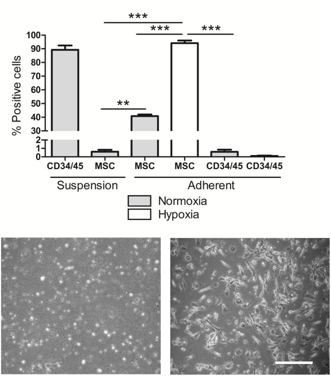
Fig 1. Peripheral blood mononuclear cell characterization.
(A) Fluorescent labelling of fresh PBMC in suspension and adherent PBMC in both normoxia and hypoxia comparing hematopoietic and mesenchymal cell surface markers (n = 4). Representative images of PBMCs after 12 days growing in (B) normoxia and (C) hypoxia (scale bar 50 μm).

