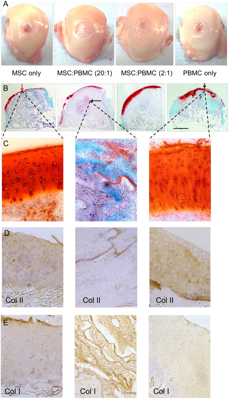
Fig 4. Analysis of the defect repair.
(A) Representative images of an average sample of each treatment group showing the macroscopic surface repair in femoral condyles. (B) Osteochondral healing of each treatment group stained with Safranin O/Fast Green. The scale bar represents the radius of the initial defect (6.0 mm). Some of the findings include: neocartilage formation on the surface of the defect (red/black vertical arrow) and remnants of the biomaterial (black horizontal arrow). (C) Safranin O/Fast Green stained high magnification (20x) images of the articular cartilage healing in the surface and in the subchondral bone where remnants of the biomaterial can be found. (D) Collagen type II staining and (E) Collagen type I staining at the repair site and within the remnants of the collagen biomaterial.

