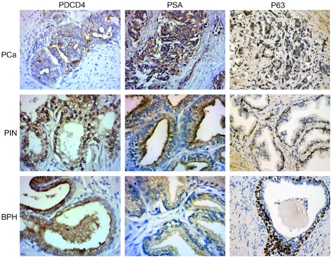
Fig 1. PDCD4 and PSA expression in prostate cancer, prostatic intraepithelial neoplasia and prostatic hyperplasia.

In prostate cancer, considerable amount of normal gland is lacking. Cancer nests are formed with irregular cribriform glands, and cancer infiltrates the muscle tissue and cell cytoplasm. This staining pattern was graded PDCD4(+), PSA(+++). In prostatic intraepithelial neoplasia, the staining pattern was graded PDCD4(++), PSA (++). In prostatic hyperplasia/glandular hyperplasia, the nuclear staining pattern was graded PDCD4 (+++), PSA (+/−). The expression of PDCD4 and PSA were different (x 2 = 8.632, P<0.05) among the 3 tissue types. In the Spearman rank test, the expression levels of PDCD4 and PSA among the 3 prostate tissue types were negatively correlated (−1 < r < 0).
