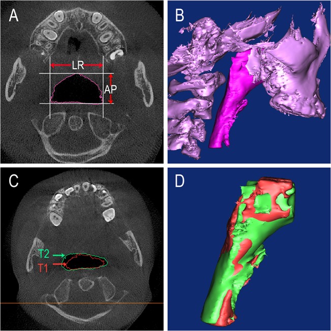
Fig 3. The changes of the upper airway.

A) The largest lateral (LR) and anteroposterior (AP) dimensions of the airway. B) The 3D model of the upper airway and maxillary in the control subjects. C) The changes of the upper airway between T1 and T2 data in theaxial view. D) After PE treatment the 3D model of the upper airway have changed.
