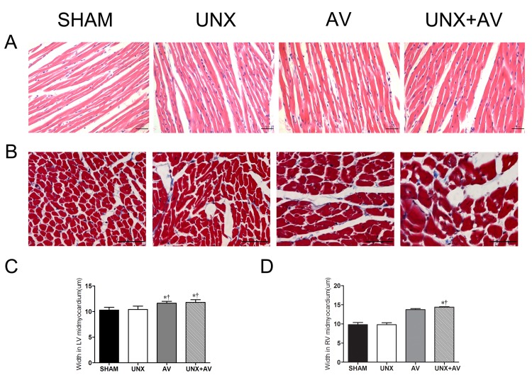
Fig 6. Histological features of cardiac tissue.
[Sham (n = 10); UNX (n = 10); AV (n = 13); UNX+AV (n = 19)].A. Hematoxylin and eosin (HE) staining, x200. B. Masson’s trichrome staining, x400. C. Bar graphs of left ventricular cardiomyocyte width. D. Bar graphs of right ventricular cardiomyocyte width. *p<0.05 vs. Sham;†p<0.05 vs. UNX;‡p<0.05 vs. AV.

