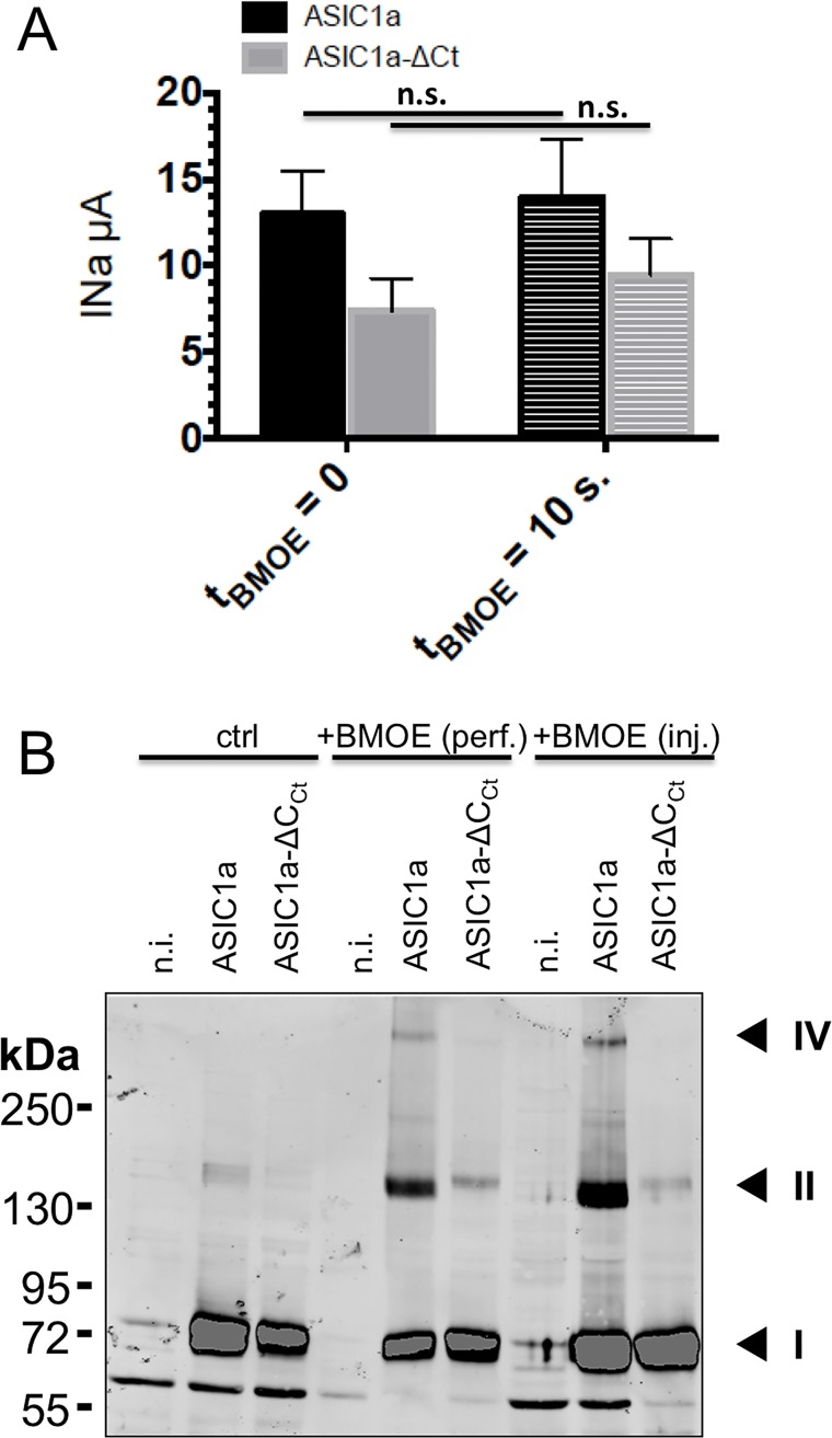
Fig 1. Effects of intracellularly applied BMOE on hASIC1a activity and oligomerization.
A: Currents were recorded in cut-open oocytes expressing either wild type ASIC1a (black bars n = 18) or ASIC1a-ΔCCt lacking cysteines in the C-terminus (grey bars, n = 17) before and after 10 s of intracellular perfusion with 2 mM BMOE (+BMOE). Bars represent mean ± SE. B: Anti-ASIC1a western from oocytes, either non-injected (n.i.), or expressing ASIC1a or ASIC1a-ΔCCt, untreated (left), or treated with 2 mM BMOE by internally perfusion (perf.) or intracellular injection (inj.). Numbers I, II, and IV designate the most prominent bands that are specific for ASIC1a, II and IV having apparent weight sizes that are, respectively, twice and four times that of I.

