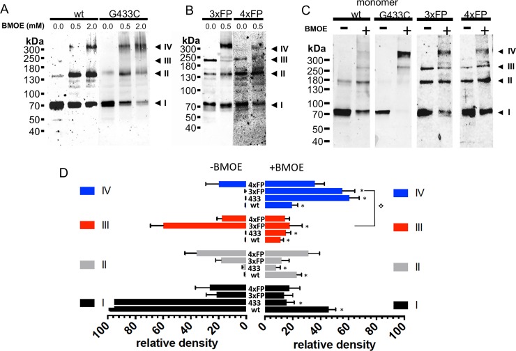
Fig 6. Oligomeric states of ASIC1a trimeric and tetrameric fusion proteins at the surface of cells expressing functional ASIC1a.
A-B: Anti-ASIC1a western blot of surface biotinylated fractions of oocytes, treated with vehicle or with BMOE (0.5 or 2 mM) before lysis, expressing either ASIC1a wt or G433C (A) or 3ASICFP (3xFP) or 4ASICFP (4xFP) (B). C: Anti-ASIC1a western blot of ASIC1a in cell-surface biotinylated fractions of CHO cells expressing monomeric ASIC1a wt, or G433C mutant, 3xFP, or 4xFP fusion proteins with or without treatment with BMOE before lysis. I, II, III, IV have the same meaning as in previous figures. D: Relative intensities (mean ±SD) of each of the 4 bands (I to IV) ASIC1a oligomers from cell-surface biotinylated fractions of Xenopus oocytes and CHO cells expressing ASIC1a (wt, n = 17), or G433C (433, n = 17) monomeric forms, or 3xFP (n = 8), or 4xFP (n = 8) fusion proteins treated with either vehicle or 0.5 mM BMOE. Symbol * denotes p<0.01 for comparison between condition -BMOE and +BMOE, ❖ p<0.01 for the indicated comparison.

