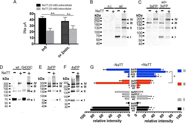
Fig 7. Oligomeric states of affinity-purified ASIC1a isolated from cells expressing functional ASIC1a trimeric and tetrameric concatemers, under oxidizing conditions.
A: Effects of extracellular (black bars) or intracellular (grey bars) perfusion of NaTT (20 mM) on ASIC1a currents measured in Xenopus oocytes before (t = 0) and after perfusion (t = 2min.). Bars represents means ± SE (n = 28). B-C: Anti-ASIC1a western blots of control or NaTT-treated (0.3 mM NaTT) affinity-purified fractions from Xenopus oocytes, non-injected (n.i.), or expressing ASIC1a (B), or 3xFP alone or co-expressed with wt ASIC1a (C). D-F: Anti-ASIC1a western blots of affinity-purified fractions from ASIC1a in CHO cells expressing either ASIC1a monomers wt or G433C mutant (D), 3xFP (E), 4xFP (F) and treated with NaTT (0.3 mM). G: Relative intensities (mean ±SD) of each of the 4 bands (I to IV) corresponding to ASIC1a oligomers identified on SDS-PAGE from cells (Xenopus oocytes and CHO cells) expressing ASIC1a wt (n = 9), G433C (n = 5), 3xFP (n = 7), or 4xFP (n = 4), without or after treatment with 0.3 mM NaTT. * denotes p<0.01

